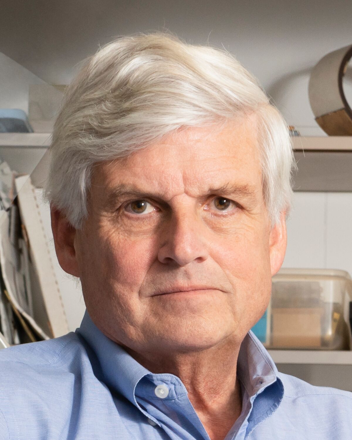
David Corey

David Corey, PhD

Bertarelli Professor of Translational Medical Science

We are interested in the gating of mechanically sensitive ion channels, which open in response to force on the channel proteins. We study these channels primarily in vertebrate hair cells -- the receptor cells of the inner ear, which are sensitive to sounds or accelerations. Hair cells are epithelial cells, with a bundle of stereocilia rising from their apical surfaces. Mechanical deflection of the bundles changes the tension in fine "tip links" that stretch between the stereocilia; these filaments are thought to pull directly on the mechanically gated transduction channels to regulate their opening.

Tip links are made of two unusual cadherins with long extracellular domains--cadherin 23 and protocadherin 15—whose N-termini join to complete the link. We are interested in the tip link’s biophysical properties and how the two cadherins join. We have determined the crystal structure of the N-termini of protocadherin 15 bound to cadherin-23, and have used steered molecular dynamics to determine the elastic properties and unbinding force of the cadherins. The crystal structures and molecular dynamics together have helped explain how deafness-producing mutations in the tip link disrupt its structure. (Sotomayor et al., 2010; 2012)

Hair bundles have an elaborate and highly stereotyped morphology, which is essential for conveying mechanical stimuli to transduction channels. As hair cells are never replaced, we are interested in how the cell maintains the bundle’s shape over months or years. Together with Claude Lechene (BWH), we tagged proteins with the 14N isotope and used multi-isotope imaging mass spectrometry to measure protein turnover. We found that stereocilia are remarkably stable, with most of their proteins lasting for months before replacement. However a half-micron zone at their tips, encompassing the transduction apparatus, shows high protein turnover (Zhang et al., 2012).

To understand the mechanics of hair cell transduction, we have characterized the movement of stereocilia bundles with high resolution light microscopy and strobe illumination. Stereocilia do not bend but pivot at their bases, and they remain touching within 10 nm even as they slide past one another by hundreds of nanometers. This “sliding adhesion” confers independent gating on transduction channels. (Karavitaki & Corey, 2010)

Transduction channels open with a mechanical stimulus, but then adapt over a time course of milliseconds. One phase of adaptation was shown to be mediated by a motor complex of myosin-1c molecules relaxing tension on the channels, but a faster phase apparently results from Ca2+ that enters through the channels immediately binding to close them. We are characterizing the site of Ca2+ action by photolytically releasing Ca2+ inside stereocilia and measuring the nanometer movements that correspond to channel closing.

A special challenge is to understand the mechanically-gated transduction channels themselves. These are most likely members of the new TMC family of membrane proteins, but much remains to be learned about the relationship between structure and function in this channel, and about the forces and movements associated with channel gating. A broad spectrum of biophysical and cell-biological methods is used to elucidate channel function.

Finally, other components of the transduction apparatus remain unidentified. High-throughput sequencing has revealed the pattern of gene expression in hair cells during development (shield.hms.harvard.edu), and these data suggest new proteins involved in transduction.

corey research

"A special challenge is to understand the mechanically-gated transduction channels themselves. These are most likely members of the new TMC family of membrane proteins, but much remains to be learned about the relationship between structure and function in this channel, and about the forces and movements associated with channel gating."

Visit here to see Dr. Corey's publications.