
Goal: We seek to understand the circuitry of the mammalian cerebral cortex and how it endows us with the ability to see . . . and hear and think and talk. Approach: We study visual cortex of alert monkeys trained to report specific aspects of their visual experience. This allows us to define the neural correlates of specific percepts and then study their underlying mechanisms by activating or inactivating components of the circuit.
Techniques: Our primary tools are extracellular electrophysiology—both with single electrodes and multi-electrode arrays—and psychophysics. They are complemented by techniques that allow us to dissect and probe cortical circuitry:
- circuit tracing with genetically modified rabies viruses
- microstimulation to insert specific signals into cortical circuits
- cortical cooling (cryoloops) to reversibly inactivate circuit elements
- multi-electrode arrays to record simultaneously from dozens of neurons
Current Projects:
- Dissecting parallel pathways. We have used reversible inactivation to show that MT neurons inherit tuning for direction and depth (binocular disparity) via independent, parallel pathways (Ponce et al. 2008). We are currently exploring the perceptual ramifications of this processing strategy (Smolyanskaya). We are also examining how these pathways interact in the context of multi-sensory integration and feature attention (Ruff).
- Probing cortico-cortical feedback. Two projects in the lab are aimed at deciphering this ubiquitous, but poorly understood, aspect of cortical connectivity. We are studying the detailed morphology of neurons that comprise the feedforward and feedback pathways (Berezovskii and Nassi). This will give us important clues as to what information the neurons integrate and how it is distributed to target areas. In order to understand the functional role of cortico-cortical feedback, we are reversibly inactivating feedback from areas V2 and V3 to V1 and then recording from V1 with multi-electrode arrays in alert monkeys (Hartmann, Trott). In collaboration with Dr. Gabriel Kreiman, we are developing computational models to account for our experimental findings and to guide future experiments (Gómez-Laberge).
The gray-level image at the top center is an autoradiograph of a section through MT cut parallel to the cortical surface. The patterns of 2dg uptake were produced by showing the animal a wide-field pattern of random dots (covering ~60 degrees of the visual field) that moved coherently at systematically varied directions and speeds while 2dg was infused intravenously. Regions of high 2dg uptake appear dark (wide-field motion columns), while regions of 2dg uptake equal to that of unstimulated cortex are lighter (local-contrast motion columns). The graphs immediately below the 2dg image depict the responses of two representative neurons to patches of random dots moving in the cellÍs preferred direction and speed as a function of the size of the random dot patch (area response test). Neurons in the dark regions respond more vigorously as the area of the random dot patch increases; neurons in the light regions respond well to small patches of motion but are indifferent to wide-field motion due to the presence of opponent surrounds.
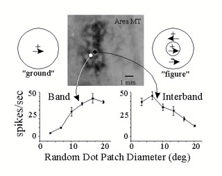 Correlation of 2-deoxyglucose (2dg) labeling and neuronal receptive field properties in the middle temporal visual area (MT).
Neuroimage
View full abstract on Pubmed
J Mater Sci Mater Med
View full abstract on Pubmed
J Neurosci
View full abstract on Pubmed
Med Image Comput Comput Assist Interv
View full abstract on Pubmed